
Lymphoma is the name for a group of blood cancers that develop in the lymphatic system. The two main types are Hodgkin lymphoma and non-Hodgkin lymphoma (NHL).

Diagnosis of Hodgkin's Lymphoma
The doctor will take your case history, carry out physical examination and recommend laboratory tests and imaging studies.
Physical examination focuses on the location and size of all palpable lymph nodes, abnormal lung sounds, and enlargement of liver, spleen, or both.
Laboratory studies
The doctor may recommend the following:
- Complete blood picture: may be normal in early stages but in advanced stages there may be either decrease in the red blood cell count (RBC) or platelet cells or white blood cell count or may be decrease in all three cells.
- Serum chemistry studies: This may show elevated calcium levels, LDH levels (an enzyme whose levels are abnormally increased in some cancers), abnormal liver function tests.
- Serum beta2-microglobulin level: May be elevated.
- HIV test
- Bone marrow biopsy is recommended, although bone marrow involvement is uncommon. Positive bone marrow will greatly influence the treatment decision.
- Imaging studies include chest x-ray, computed tomography (CT), positron emission tomography (PET)
Classification and staging of Hodgkin’s lymphoma
The diagnostic tumour cells are Hodgkin-Reed-Sternberg (H-RS) cells. The classification is based on the number and appearance of H-RS cells into 4 subtypes:
- Nodular Sclerosis
- Mixed Cellularity
- Lymphocyte-Depleted
- Lymphocyte-rich
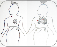
Staging: Ann Arbor staging classification is currently used along with pathology to guide the treatment choice for both Hodgkin’s and non-Hodgkin’s lymphoma.
Ann Arbor Staging Classification.
Stage I: Involvement of a single lymph node region or lymph node structure (eg, spleen, thymus or Waldeyer’s ring)
Stage II: Involvement of two or more lymph node regions on the same side of the diaphragm
Stage III: Involvement of lymph node regions on both sides of the diaphragm
Stage IV: Disseminated (multifocal) involvement of one or more extralymphatic organs, with or without associated lymph node involvement; or isolated extra lymphatic organ involvement with distant (non regional) nodal involvement
Diagnosis of Non-Hodgkin's Lymphoma
Examination in patients shows swollen spleen and liver and enlargement of lymph nodes
Laboratory investigations
The doctor may suggest the following tests.
- Complete blood picture: May be normal in early-stage disease; in more advanced stages, may demonstrate decreased RBC count (anaemia), decreased WBC count (leucopenia), decreased platelet count (thrombocytopenia).
- Serum chemistry studies: May show elevated calcium levels, abnormal liver function tests
- Serum beta2-microglobulin level: May be elevated
Imaging
The following imaging studies should be obtained in a patient suspected of having NHL:
- Chest radiography
- CT scanning of the neck, chest, abdomen, and pelvis
- PET scanning
- Bone scanning: Only in patients with bone pain, elevated alkaline phosphatase, or both
- MRI of brain/spinal cord: For suspected primary brain lesions or tumors, lymphomatous meningitis.
- Biopsy: The diagnosis of NHL depends on pathologic confirmation following appropriate tissue biopsy. The following are procedures in cases of suspected NHL:
- Bone marrow aspiration and biopsy
- Excisional lymph node biopsy
- Lumbar Puncture (A procedure in which a needle is inserted into the lower part of spine to collect cerebrospinal fluid)
Staging: Ann Arbor Staging Classification
Stage I: Involvement of a single lymph node region or lymph node structure (eg, spleen, thymus or Waldeyer’s ring)
Stage II: Involvement of two or more lymph node regions on the same side of the diaphragm
Stage III: Involvement of lymph node regions on both sides of the diaphragm
Stage IV: Disseminated (multifocal) involvement of one or more extralymphatic organs, with or without associated lymph node involvement; or isolated extra lymphatic organ involvement with distant (non regional) nodal involvement
Diagnosis of Burkitt's Lymphoma
The doctor may suggest some of the following:
- Computed tomography (CT) imaging of the chest, abdomen, and pelvis
- Chest X-ray, PET or gallium scan (looks for inflammation and uses a material called gallium)
- Bone marrow biopsy, examination of spinal fluid
- Blood tests to measure kidney and function
- Testing for HIV
- Biopsy (if Burkitt’s lymphoma is suspected, all or part of an enlarged lymph node or other suspicious disease site will be biopsied)
Staging: Ann Arbor Staging
Stage I: The lymphoma is only in one group of lymph nodes in one particular area of the body.
Stage II: More than one group of lymph nodes is affected, but all the affected nodes are contained within either the upper half or lower half of the body.
Stage III: Lymphoma is present in lymph nodes in both the upper and lower parts of the body (above and below the diaphragm). The spleen is considered as a lymph node in this staging system.
Stage IV: The lymphoma has spread beyond lymph nodes to other organs, i.e to sites such as the nervous system, bone marrow, liver or lungs.
Changed
10/Jan/2019
Community
Condition