
The number of people (aged 40-80 years) with glaucoma worldwide was estimated at 64.3 million in 2013, and is expected to increase to 76.0 million in 2020.
It is an eye disease which is one of the leading causes of blindness worldwide. It is often referred to as a “silent killer” as it does not manifest any symptoms until extensive peripheral visual loss becomes apparent in the final stages of the disease.
What does a complete glaucoma evaluation include?
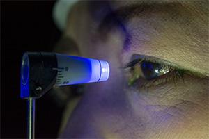
A comprehensive eye examination includes screening tests for glaucoma. A simple, painless procedure called tonometry measures the internal pressure of the eye. Intraocular pressure in most individuals ranges between 10 and 20 mmHg. A characteristic deterioration of the optic nerve associated with cupping and atrophy is common denominator to all forms of glaucoma (primary or secondary, open or closed angle, chronic or acute). Recognition of these changes in the optic nerve by opthalmoscopy is an important tool for glaucoma screening.
A complete glaucoma evaluation is indicated if a patient has an Intraocular pressure above 20 and/or suspicious looking optic nerve on a regular eye examination.
Once glaucoma is suspected the patient undergoes a thorough evaluation. A complete medical and ocular history and examination helps in diagnosing the type of glaucoma.
 - Using a variety of gonioscopic lenses, the peripheral iris, cornea and trabecular meshwork are directly visualized to determine the presence of angle closure, adhesions, inflammatory foci, traumatic injury, masses or other lesions. This helps in diagnosing the type of glaucoma and outlining the management.
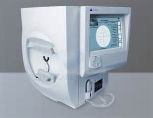
Visual Field Analysis -The functional status of the optic nerve can be assessed by specialized testing of the peripheral vision, the "visual field.” Progressive loss of the optic nerve fibers leads eventually to progressive loss of visual field and finally to complete loss or blindness. Early peripheral visual field loss is not noticeable to the patient, and its slow progression makes its recognition nearly impossible without this special testing. Early detection is therefore important to limit the permanent damage. Also assessment of visual field changes on follow up helps to monitor the progression of glaucoma on treatment.
Optical Coherence Tomography (OCT) - is a relatively newer imaging modality that detects early disease and progression in glaucoma suspects. It may help in diagnosing glaucoma even before visual fields get affected. SD-OCT identifies and monitors structural damage by measuring peripapillary ( around the disc) RNFL thickness.
Changed
04/Nov/2017
Condition