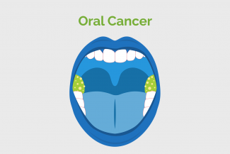
Oral cancer refers to cancers of the head and neck. It includes cancer of the lips, tongue, cheeks, gums, salivary glands, floor of the mouth, hard and soft palate, sinuses and pharynx. Brain cancer falls in a different category.
The doctor may recommend some of the following tests:
Colposcopy and optical diagnostic systems: The colposcope is an instrument with a magnifying lens that allows the doctor to take a close look at the affected part of the mouth. Acetic acid solution and iodine solution (Lugol's or Schiller's) are applied to the surface to improve visualisation of abnormal areas.
Toluidine blue (TB) and lugols iodine staining: is a simple and inexpensive diagnostic tool to highlight abnormal areas of mucosa. Toluidine blue is applied to the suspected area and the patient is asked to wash the mouth. The stain will adhere to the abnormal area and help the doctor identify the area from where he should take the biopsy. Lugols iodine works in a similar way. Abnormal tissue stains brown while normal tissue does not.
Exfoliative Cytology: This is done for patients who are asymptomatic or have minor symptoms that do not warrant a biopsy. The suspicious area is scraped for surface cells that are sent for lab investigation.
Biopsy: A small sample of tissue from the suspicious area is taken and sent to the lab for investigation. There are different types of biopsy:
- Fine-needle aspiration: A long, thin needle is inserted into the suspicious area. A syringe is used to draw out fluid and cells for examination.
- Core needle biopsy: A larger needle with a cutting tip is used during core needle biopsy to draw a column of tissue out of a suspicious area.
- Excisional Biopsy: A whole organ or a whole lump is removed
- Incisional biopsy: Only a portion of the lump is removed surgically.
- Vacuum-assisted biopsy: A suction device increases the amount of fluid and cells that are extracted through the needle. This can reduce the number of times the needle must be inserted to collect an adequate sample.
- Image-guided biopsy: Image-guided biopsy combines an imaging procedure such as X-ray, computerised tomography (CT), magnetic resonance imaging (MRI) or ultrasound with a needle biopsy.
Plain film radiographs (X-rays of the mouth): Radiographs (X-rays) check whether the cancer has extended into the bone.
Scans: CT, MRI, PET- CT can assist in the diagnosis and to see if the cancer has spread.
Changed
16/Jan/2019
Community
Condition